
0SPK34Z is a medical procedure where a surgeon removes a device used to stabilize the right tarsometatarsal joint through a minimally invasive approach. This is typically done to address any complications or to allow for proper healing of the area.
Table of Contents:
- 🔎 Clinical Indication
- 📋 Preparation
- 📖 Methodology
- 🩹 Recovery
- 🚨 Complexity & Risk
- 🔀 Similar Procedures
🔎 Clinical Indication
0SPK34Z, the removal of an internal fixation device from the right tarsometatarsal joint, may be necessary if the device is causing pain, discomfort, or complications. This procedure is typically performed through a minimally invasive percutaneous approach to reduce the risk of infection and minimize recovery time.
Internal fixation devices such as screws or plates are commonly used in orthopedic surgeries to stabilize bones during the healing process. However, these devices may need to be removed once the bone has fully healed to prevent irritation or issues with movement. The decision to remove an internal fixation device will depend on the individual’s symptoms and the recommendation of the healthcare provider.
Overall, the removal of an internal fixation device from the right tarsometatarsal joint is a routine procedure that aims to improve the patient’s comfort and function. By addressing any issues related to the device, patients can experience relief and potentially regain full mobility in the affected joint.
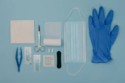
📋 Preparation
Before undergoing 0SPK34Z, the patient will need to have a comprehensive evaluation by their healthcare provider. This will involve a physical examination and review of medical history to ensure they are a suitable candidate for the procedure.
In addition, the patient may need to have imaging tests such as X-rays or CT scans to help the surgeon plan the procedure and identify the exact location of the internal fixation device. This will ensure that the device is removed safely and effectively.
The patient may also be instructed to fast for a certain period before the procedure and to follow specific pre-operative instructions provided by their healthcare team. This will help minimize any potential risks and ensure a successful outcome for the removal of the internal fixation device from the right tarsometatarsal joint using a percutaneous approach.
📖 Methodology
During 0SPK34Z, a surgeon uses a minimally invasive technique to remove hardware from the right tarsometatarsal joint. This hardware may have been used to stabilize fractures or correct deformities in the foot.
The percutaneous approach involves making small incisions through which instruments are inserted to remove the internal fixation device. This method typically results in less scarring and a quicker recovery time for the patient.
🩹 Recovery
After the removal of internal fixation device from the right tarsometatarsal joint using a percutaneous approach, the patient will typically experience some discomfort and swelling in the area. This is a normal part of the recovery process as the body heals from the procedure.
The patient may be prescribed pain medication to manage any discomfort post-operation and advised to keep the foot elevated to reduce swelling. Physical therapy may also be recommended to help restore strength and mobility in the joint.
Overall, the recovery process for this procedure is usually relatively quick, with most patients able to return to normal activities within a few weeks. It is important for the patient to follow their healthcare provider’s instructions carefully to ensure a smooth and successful recovery.
🚨 Complexity & Risk
Performing 0SPK34Z, or the removal of an internal fixation device from the right tarsometatarsal joint using a percutaneous approach, is a complex procedure. The tarsometatarsal joint is a critical joint in the foot that is responsible for weight-bearing and movement, making the procedure delicate and requiring precision.
One potential risk for patients undergoing this procedure is infection. The process of removing the internal fixation device can introduce bacteria into the surgical site, increasing the risk of infection. Patients should be closely monitored for any signs of infection post-surgery, such as increased pain, swelling, or redness at the surgical site.
In addition to the risk of infection, patients may also experience complications such as nerve or blood vessel damage during the procedure. The small size and intricate nature of the tarsometatarsal joint make it challenging to navigate during surgery, increasing the likelihood of accidental damage to surrounding structures. Patients should be informed of these potential risks before undergoing the procedure.
🔀 Similar Procedures
Another medical procedure similar to the Removal of Internal Fixation Device from Right Tarsometatarsal Joint, Percutaneous Approach is the Removal of Cranial Fixation Device from Skull, Percutaneous Approach. This procedure involves removing hardware, such as screws or plates, from the skull through a minimally invasive approach.
Both procedures aim to remove internal fixation devices that were previously implanted to stabilize bones or joints. By removing these devices, patients may experience decreased pain and improved range of motion in the affected area. These procedures are typically performed under local anesthesia and have a relatively quick recovery time.